


John Tolley, February 8, 2020
The University of Illinois has a laser-focus on improving cancer pathology, quite literally.
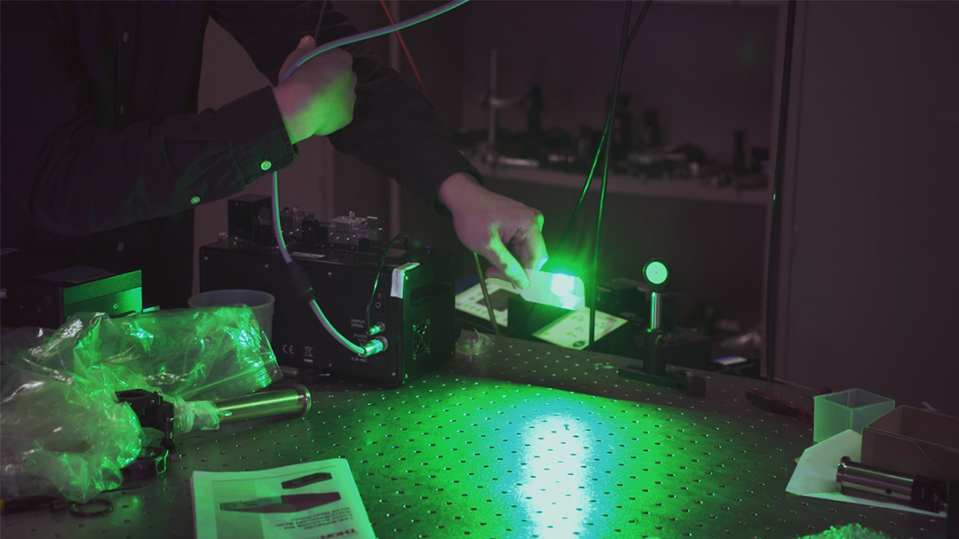
Laser microscopy techniques under development at the Carle Illinois College of Medicine have the potential to allow doctors to detect cancers earlier and in enhanced detail.
?In the Biophotonics Imaging Lab, we develop novel optical imaging and sensing technologies,? explains lab director Dr. Stephen Boppart, MD, PhD, the Abel Bliss Professor of Engineering. ?We know in cancer there?s a lot that goes on at the very early level when cells mutate and become cancerous spread. What we want to do is be able to image at that scale. To be able to catch disease in cancer when it?s just starting instead of it growing to be a large mass then that?s later detected on CAT scans or MRIs.?
For well-over a century, the diagnosis of cancer has relied on the biopsy. This procedure removes a sample of suspected cancerous cells from the body for testing.
To varying degrees, biopsies are invasive, and they have intrinsic downsides, says Sixian You, a graduate researcher in the Biophotonics Imaging Lab.
?[The sample] goes into a huge machine where it gets stained,? You explains. ?It goes through all these processes to get to a point where you can see them in the microscope. And this whole process takes quite a few hours and it kills tissue before it sees the tissue. So, we don?t have the living information anymore, which is actually very important for cancer diagnosis?
Another prominent drawback to biopsies is the time it takes for extraction, treatment of the sample, and lab analysis. For critical situations, where time is of the essence, this delay in diagnosis can severely impact outcomes.
Boppart and his team are essentially removing the middleman in cancer pathology. Instead of staining an extracted sample to highlight contrast between various structures and tissues, their method uses lasers and optic fiber. The detail afforded by the laser imaging allows them to see a wide-spectrum of tissues, cells and cell anomalies.
?What we?re trying to do though is really make this better for the patient, to be able to diagnose disease early and give them information so they don?t have to wait for that diagnosis,? says Boppart. ?What we want to do is make microscopes that can be used right during the procedure, during surgery or in the office and so that the surgeon or the doctor is going to be able to see the tissue at that scale and perhaps even make a diagnosis.?
In addition to first-line cancer pathology, the technology also has utility in surgical settings. The team is concurrently developing compact, portable laser microscopy tools for use during tumor removal surgeries. This could help surgeons more thoroughly identify and remove all cancerous tissues.
?The goal is really to have a diagnosis quickly, at the point of care, so the surgeon knows, do I need to take more tumor out or am I done,? says Boppart.
The unique structure of the Carle Illinois College of Medicine makes possible advancements such as this. As the first engineering-focused medical school in the nation, Carle brings together a variety of researchers to drive medical innovation in the 21st century.
Boppart also credits Illinois? Beckman Institute for Advanced Science and Technology, which focuses on Intelligent systems, integrative imaging, and molecular science and engineering, as an engine of interdisciplinary collaboration.
?This is a very special place where faculty and students and researchers from all across campus, many different fields, come together to try to solve the complex problems that no one field could solve on its own,? Boppart remarks. ?So, being here at this Beckman Institute, it?s been wonderful because we?ve been able to work with colleagues in chemistry and life sciences and computer science, engineering, medicine, we all come together to try to tackle these types of problems.?