


John Tolley, December 8, 2018
The islets of Langerhans, the corpus callosum, Glisson?s capsule, Auerbach?s plexus. These colorful names highlight the diversity of tissues that constitute our bodies. They carry out myriad functions, from regulating our digestion to filtering impurities in our blood to enjoining the hemispheres of our brain.
But these tissues, for all of their profound specialization, are intensely intricate, infinitesimal and tucked deep within our bodies. To study their form and function - or, malfunction as it may be - would seem to require scientific sorcery worthy of Fantastic Voyage. Unless it didn?t.
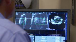
Modern clinical and hospital MRI scanners offer a glimpse beneath the surface of our running systems, but at an average maximum field strength of 3 tesla, they?re scope of view is limited, relatively speaking. A ground-breaking new MRI scanner at the University of Minnesota, however, is set to provide doctors and researchers with previously unattainable levels of clarity and resolution in the imaging of the body.
?Here at the University of Minnesota, we designed and developed the 10.5 tesla MRI system,? explains Dr. Greg Metzger, an associate professor at the Center for Magnetic Resonance Research (CMRR) in the University of Minnesota Department of Radiology. ?This scanner consists of over a thousand kilometers of niobium-titanium superconducting wire, which is bathed in over 4,500 liters of helium. This scanner, which weighs 110 tons, is shrouded in a steel shield of over 600 tons to shield its fringe fields from the rest of the center. This scanner, we hope, will give us increased sensitivity and resolution for furthering our understanding of brain function and connectivity.?
Driven by the limitations of more common systems, the University of Minnesota has over 25 years of experience in developing cutting-edge high-field MRI systems. The newly developed 10.5 t model, which is currently undergoing safety testing in conjunction with the FDA, will allow clinicians to study carbon, phosphorous and sodium levels in cell nuclei, brain energetics, treatment progression, and even the biomechanics of aging.
Another intriguing field of research that has the potential to be more fully elucidated with the powerful device is mental illness, notes Dr. Kamil Ugurbil, McKnight Presidential Endowed Chair professor of radiology and director of the CMRR.
?Neuro-psychiatric disease is the number one source of disability in the United States; it's not cancer, it is not heart disease, it is neuro psychiatric diseases,? says Ugurbil. ?Yet, we don't understand these diseases very well. With this high magnetic field, we can study brain anatomy, brain structure, brain activity with much higher resolution. That means that we can get information from even smaller pieces of tissue in the brain, and we can study it much more accurately.?
The University of Minnesota has a long history of medical technology innovation, frequently partnering with numerous Minnesota-based biotech firms to advance research in the field. As Metzger explains, this is a key factor in the development of next-generation MRI scanners.
?Because of the support from the state, the university and National Institutes of Health, we have a very unique environment here which allows us to push field strength and its use in clinical and basic research. We have a unique set of personnel that allow us to push the fields and develop the technologies necessary to exploit the advantages of high field.?